loading...

by Tim Cassidy | Apr 7, 2025 | Uncategorized
Online CE Credits is thrilled to announce the launch of a groundbreaking new course, “EMDR 2.0 Skills Mastery: Techniques and Practical Applications,” designed to empower therapists with cutting-edge tools to enhance trauma therapy. This course, now available for enrollment, offers mental health professionals an opportunity to deepen their practice and deliver faster, more effective care to clients.

Building on the proven foundation of traditional EMDR (Eye Movement Desensitization and Reprocessing), EMDR 2.0 introduces advanced techniques to accelerate and deepen the processing of traumatic memories. Key enhancements include working memory taxation techniques and client buy-in strategies. These methods, informed by the latest neurobiological research, aim to provide nuanced, impactful treatment for clients with complex trauma.
“EMDR 2.0 represents a significant step forward in trauma therapy,” says Dr. Jennifer Sweeton, course instructor and a renowned trauma psychologist. “We’re equipping therapists with practical, evidence-based strategies to help clients heal more efficiently, even in challenging cases.”
The live webinar is on January 30, 2025, and afterward will be accessible on demand 24/7, offering flexibility for busy professionals. Therapists can earn continuing education credits while learning at their own pace. Online CE Credits encourages clinicians to stay ahead in their field by integrating these innovative techniques into their practice.
For more information and to register, see: https://onlinececredits.com/events/emdr-2-0-advanced-techniques-for-fast-safe-effective-treatment/

by Tim Cassidy | Mar 10, 2025 | Educational, Uncategorized
Introduction
For mental health professionals, understanding the brain can unlock powerful insights into client care. In The Neuroscience Handbook for Clinicians by Dr. Jennifer Sweeton, the thalamus emerges as a critical, often overlooked brain region in trauma therapy. As a “relay station” for sensory information, the thalamus plays a pivotal role in how clients experience PTSD symptoms like re-experiencing and dissociation. Let’s explore its functions, impact on trauma, and how you can apply this knowledge in your practice, and how our CE courses at Online CE Credits can help.
What Is the Thalamus and Why It Matters
The thalamus, a small egg-shaped structure above the brain stem, acts as the brain’s sensory gatekeeper, relaying sensory inputs (except smell) to regions like the amygdala (threat detection) and cortex (executive functioning). That sensory information reaches the amygdala in just 12 milliseconds, which is half the time it takes to reach the cortex (25 milliseconds). This means clients often process danger before rational thought kicks in, a key factor in trauma responses.
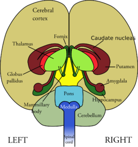
The Thalamus in Trauma: Symptoms and Challenges
Research (e.g., Etkin & Wager, 2007; Kim et al., 2007) shows that individuals with PTSD often exhibit thalamic hypoactivation at rest and during re-experiencing symptoms, such as traumatic sensory re-enactments. This reduced activation disrupts sensory filtering, making trauma memories feel vivid and present, as if the event is happening now. And, there is weaker connectivity between the thalamus and amygdala in severe PTSD, exacerbating these symptoms (Zhu et al., 2017). Conversely, during dissociation or flashbacks, the thalamus becomes hyperactive, over-filtering sensory input, leading to numbness or disconnection. When this happens, clients may appear “spacey” or detached, per Lanius et al. (2001).
Applying Neuroscience to Trauma Therapy
Understanding the thalamus’s role can transform your approach. Research has shown that Eye Movement Desensitization and Reprocessing (EMDR) activates the thalamus through bilateral eye movements, helping clients better filter sensory information and integrate traumatic experiences (Bergmann, 2008). This reduces the intensity of re-experiencing symptoms by balancing internal and external awareness. Cognitive Behavioral Therapy (CBT) also shows promise, increasing thalamus activation and reducing PTSD symptoms, per Peres et al. (2007). These brain-based strategies can guide your treatment planning, ensuring client-centered care grounded in neuroscience.
Earn CE Credits with Online CE Credits
At Online CE Credits, we offer NBCC- and ASEB-approved courses to deepen your neuroscience-based practice. With over 250 on-demand courses, you can learn 24/7, earning credits at your pace. Explore our trauma-focused CE programs and start transforming your practice today!
Conclusion
The thalamus’s role in sensory processing makes it a key player in trauma therapy. By understanding its impact on PTSD symptoms and leveraging therapies like EMDR and CBT, you can help clients heal more effectively. Explore these insights further with Online CE Credits and take your practice to the next level.
by Tim Cassidy | Mar 10, 2025 | Educational
Introduction
Ever wondered how brain science can enhance your therapy practice? At Online CE Credits, we believe understanding the brain can transform client care. Let’s explore how our CE courses can empower you with brain-based strategies to use with your clients!
Breaking Down Neuroscience for Clinicians
There are eight key brain regions (e.g., thalamus, amygdala, hippocampus) involved in mental health, linking their changes to symptoms like anxiety, trauma, and addiction. For example, the thalamus, a sensory relay station, underactivates in PTSD, intensifying re-experiencing symptoms by failing to filter sensory inputs, per Etkin & Wager (2007). Understanding these connections helps you identify why clients behave as they do in the therapy room.
Elevate Your Skills with Online CE Credits
At Online CE Credits, we bring neuroscience to life with over 250 accredited courses, including neuropsychotherapy training. Learn how brain regions like the insula and amygdala impact therapy, start applying brain science to your practice, and earn CE credits at your pace, 24/7!
Conclusion
Neuroscience doesn’t have to be daunting. With Online CE Credits, you can integrate brain-based insights into your practice, helping clients heal more effectively!

by Tim Cassidy | Mar 10, 2025 | Educational
Introduction
Anxiety affects millions of people, and understanding the brain science of anxiety can revolutionize your therapy approach. In The Neuroscience Handbook for Clinicians by Dr. Jennifer Sweeton, the thalamus, a sensory relay station, emerges as a key player in anxiety disorders. At Online CE Credits, we help therapists apply brain science to client care. Let’s dive into the thalamus’s role in anxiety and how you can use this knowledge to earn CE credits and enhance your practice.
The Thalamus’s Role in Anxiety
Located above the brain stem, the thalamus relays sensory information to regions like the amygdala. In anxiety, the thalamus, particularly its paraventricular nucleus (PVT), facilitates fear conditioning, per Wilensky et al. (2006). This means that anxious clients learn fear faster due to PVT activation, amplifying sensitivity to threats, per Penzo et al. (2015). Thalamic activation also spikes during anticipatory anxiety (e.g., awaiting a phobic stimulus, per Straube et al., 2007) and worry in generalized anxiety disorder (GAD), per Karim et al. (2017), presenting as jumpiness, vigilance, or dread.
Practical Implications for Therapy
Clients with anxiety may seem “primed” for fear, reacting intensely to perceived threats due to thalamic involvement. However, the good news is that therapies like CBT reduce thalamic activation, lowering reactivity to anxiety cues, per Furmark et al. (2002). Medications (e.g., paroxetine, fluoxetine) also decrease thalamic connectivity, easing anticipatory anxiety, per Duval et al. (2015). Use this insight to select CBT techniques (e.g., cognitive restructuring, exposure) that target fear processing, helping clients manage worry and reduce vigilance.

Deepen Your Expertise with Online CE Credits
At Online CE Credits, several of our accredited CE courses focus on the treatment of anxiety, offering actionable strategies for anxiety treatment. Earn credits 24/7 at onlinececredits.com!
Conclusion
The thalamus’s role in fear conditioning and anticipatory anxiety offers a brain-based lens for therapy. Apply these insights with CBT, and deepen your knowledge with Online CE Credits courses!

by Tim Cassidy | Mar 10, 2025 | Educational
Introduction
Anxiety treatment can be complex, but understanding the brain can simplify your approach. The amygdala, known as the brain’s “fear center,” plays a starring role in anxiety disorders. Let’s explore the amygdala’s impact on anxiety and how you can earn CE credits to enhance your practice.
The Amygdala: Anxiety’s Fear Center
Located in the temporal lobes, the amygdala is the brain’s fear processing hub. It identifies threats, drives fear learning, and regulates stress responses, per Abuhasan & Siddiqui (2018). In anxiety disorders, the amygdala becomes hyperactive, reacting intensely to perceived dangers, per LeDoux (2007). The left amygdala tends to process positive emotions (e.g., happiness, per Lanteaume et al., 2007), but its right side dominates in anxiety, fueling negative emotions and risk-taking, per De Martino et al. (2010). This hyperreactivity manifests as vigilance, jumpiness, or dread in clients.

Using Neuroscience in Anxiety Therapy
The amygdala’s role suggests targeting fear processing in therapy. Exposure therapies (e.g., Prolonged Exposure, EMDR) can reduce amygdala activation, helping clients manage fear responses. Somatic and mindfulness practices can also lower amygdala reactivity, fostering emotional regulation, per MacNamara et al. (2016). Combine these with CBT to strengthen amygdala connectivity with the prefrontal cortex, further reducing anxiety symptoms.
Conclusion
The amygdala’s role in fear processing offers a brain-based lens for anxiety therapy. Apply exposure and mindfulness techniques to reduce its reactivity, and deepen your expertise with Online CE Credits at onlinececredits.com. Join 40,000+ professionals, and elevate your practice now!